Elastography - accents
Many diseases, predominantly malignant, cause changes in the mechanical properties of tissues. These changes cannot be directly measured in CT and traditional ultrasound.
Elastography is an imaging technique that increases the specificity of detection of malignancies from 85% in conventional ultrasound to 98.5% using elastography.
Elastography of the mammary gland can significantly improve the ability of echomammography to distinguish between benign and malignant lesions of the breast. This reduces unnecessary biopsies.
Elastography
Mammography and ultrasound examination of the mammary glands is often used in combination to characterize tumor formations and to assess the risk of malignancies. Ultrasound elastography has been introduced as an additional method for improving the characterization of lesions.
The examination begins with a high-frequency ultrasound scan in B-scan mode, and then elastography is applied when it is clinically necessary to distinguish malignant susceptibility lesions, which improves the accuracy of BI-RADS classification. Elastographically, lesion rigidity can be assessed by mapping tissues subjected to external pressure through the exploratory ultrasonic sensor.
There are different technical approaches for elastography: /USE/freehand elastography/, SWE/shear-wave elastography/, the newest one ARFI/acoustic radiation force impulse/- acoustic impulse technology, and SSI/supersonic shear-wave imaging/ - image obtained by ultrasound wave.
Elasticity Coefficient, measured in kPa, is used for tissue evaluation, which for normal fibro-glandular tissue is 28 to 66 kPa, fatty tissue is 18-24 kPa, and for carcinomas, it reaches 560 kPa. A histogram (Strain-Histogram) and lesion staining can visualize the area of tumor infiltration and even invisible to the B-mode carcinomas.
What is Elastography examination?
Compression elastography is based on the application of mild breast pressure and the measurement of the form-deformation effect, thus providing the value of the lesion's hardness to surrounding tissues. The technique only allows qualitative and semi-quantitative analysis by calculating the deformation factor rather than the absolute elasticity. The application of elastography is in the addition of important details and the determination of epithelial and connective tissue components by their different elasticity, and technology is particularly useful in focal pathologies. Benign forms are usually hypoechogenic modules, more often in regular shape and transparent borders, oval with longitudinal orientation parallel to the skin. Generally, benign lesions are more rigid than healthy tissue but softer than malignant tumors, and they make hyalinated fibroadenoma and fatty necrosis sites.
Benign lesions are described in elastography as lesions with a low score on the color map (score 1-2), while the malignant have a score above 3-4. Fibroadenomas with a large fibrous component and reduced water content may have a dubious look on the color map but in any case lower density than malignant lesions.
Fat necrosis is usually visualized as a hypoeogenic band with non-homogenous fibrosis formation. Evolution to sclerosis may cause images to be controversial in the differential diagnosis. The sclerosing lesion causes architectural disturbances of the normal glandular structure by overlapping the fibrous reaction of the surrounding breast tissue and increasing their hardness which is elastically distinguished as a tissue with a higher hardness than normal tissue but lower than that of malignant lesions and so becomes distinct. Fat necrosis is reliably diagnosed by mammography, while radial lesions remain a diagnostic challenge, which, if it can not be differentiated, is elastographycally diagnosed by biopsy. The purpose of elastography is to reduce the number of unnecessary biopsies.
Malignancies are ascertained with the presence of acoustic shadow, hyperechogenic aureole, desmoplastic lesion reaction, calcifications, dilation of the milk ducts, glandular edema, and significant vascularization. Carcinomas are less susceptible to deformation than the test pressure compared to normal tissues, which can quickly diagnose them elastographically. Estimates of the elasticity of the carcinomas and metastatic lymph nodes are usually represented as blue areas in the color map. Smaller malignant lesions such as medullary, mucous, papillary, and necrotic ductal carcinoma are rare. Ecomagraphy has higher sensitivity than mammography, which has limited diagnostic value and sensitivity - only 48% in patients with dense mammary glands. When combining the two methods, the sensitivity increases to 97%.
Elastography increases the specificity of the conventional B-scan mode to 98.5%, which helps diagnose malignant lesions with an atypical appearance in B-mode. This improves accuracy when inserting the lesion into a different BIRADS category. For example, a tumor formation that is classified as a BIRADS 3-possibly benign lesion, by elastography can be refined and placed in the next BIRADS 4-Suspected Malignant Lesion category, in the presence of evidence of increased lesion and stain hardness and in blue, or to lower the inferior BIRADS 2-benign lesion group if elastographic is benign color mapping. The elastoclastic classification Tsukuba Elasticity Score is 1 and 2 benign, 3 is likely benign and 4 and 5 malignant lesion. The cysts there is BGR-Sign.
Elastography can also be used in the diagnosis of thyroid carcinomas and metastatic lymph nodes in the neck and mammary glands. As a superficial structure as well as in the elastography of the mammary glands, the difficulties associated with obtaining a quality image, such as in the liver elastography and the deeply located lesions, are avoided. In elastography of the thyroid gland, the coloration is green in score 1 to blue in score 4.
Elastography - contact us
For additional information for the team, that makes the specialized examination at Dr. Shterev Hospital, and to book your appointment, call our Contact Center at +359 2 920 0901. Our coordinators in Sector Patients Care will answer all your questions.
Video: Gynecological and mammographic prophylactic at Dr. Shterev Hospital
Prevention saves lives! This is the credo of the Preventive Medicine Sector at Dr. Shterev Hospital. To underline the importance of medical prevention for women’s health, we created an exclusive video in which we present essential aspects of gynecological and mammalogical prophylactic care. Watch the video to learn about the latest trends in female prophylaxis, and remember that your health is in your hands.

Elastography - resume
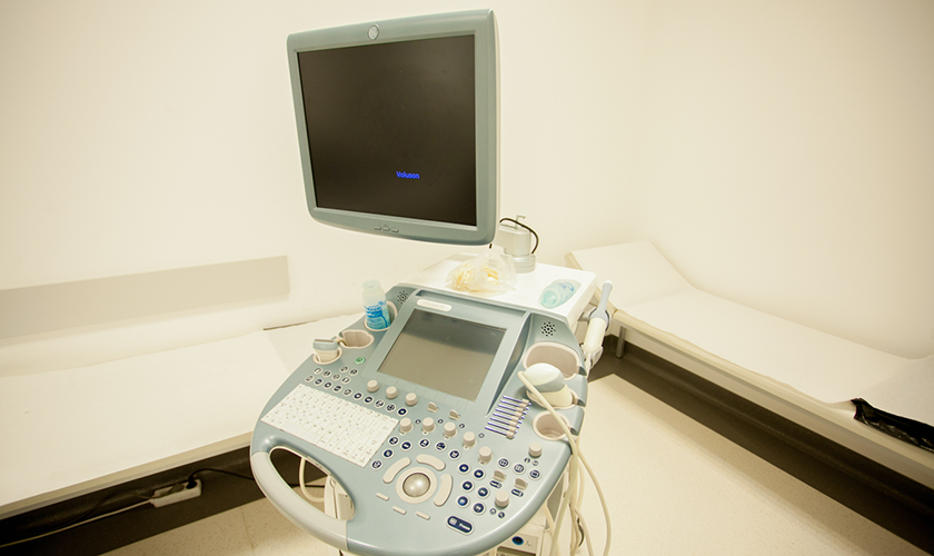
Elastography is an additional option for high-quality B-regime of scanning for diagnosis of high-density lesions, such as malignancies. It has proven reliability in distinguishing between benign and malignant lesions, improving the specificity and accuracy of ultrasound research. Dr. Shterev Hospital disposes of the necessary equipment and expertise of the medical team for conducting the specific study under all European standards.