Fetal echocardiography - accent
- The optimal term for fetal echocardiography is between the 18 and 22 weeks of gestation. At this term, the heart of the fetus is large enough to evaluate its structures.
- The required time for the procedure is about 30-45 minutes, and no special preparation is required.
- "A large percentage of the possible heart abnormalities can be detected before the baby was born may be diagnosed when the heart is evaluated by fetal echocardiography. This is because all heart’s structures are explored in detail in fetal echocardiography," Dr. Natalia Eftimova, fetal cardiology specialist.
Fetal echocardiography
What is fetal echocardiography?
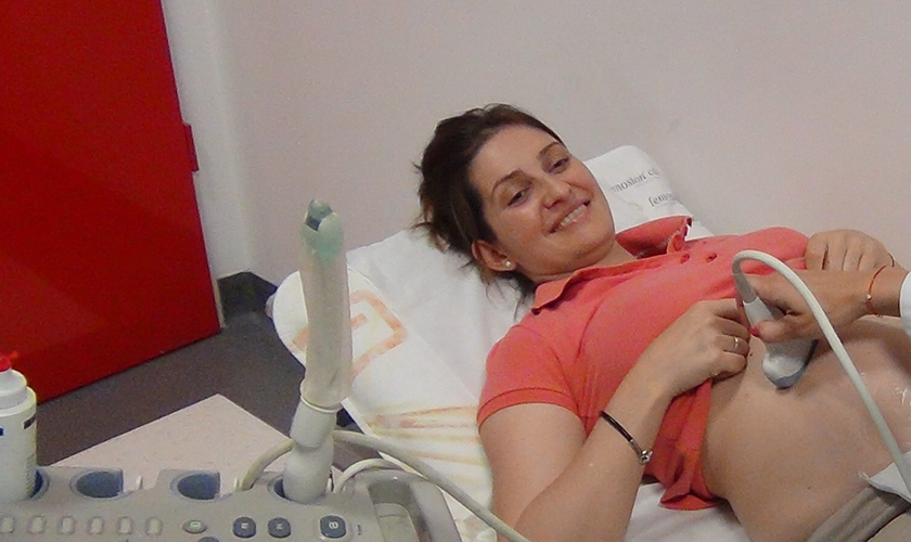
Fetal echocardiography is a specialized ultrasound examination done during the pregnancy by a pediatric cardiology specialist. Fetal echocardiography examines the position, size, structure, function, and rhythm of the baby's heart in the womb. The scan uses two-dimensional echocardiography (2D), Doppler for heart rate and vessel blood flow assessment, color-coded Doppler, visualizing different monitor colors to accurately determine the direction of blood flow and complementing Doppler's study. In some instances, the results of the fetal echocardiography determine the time for delivery, as well as the way of birth - either normal or operative. Prenatal diagnosis of congenital heart abnormalities could prevent some of the complications associated with late diagnosis.
When is fetal echocardiography recommended?
The scan is recommended in cases with abnormalities in the fetus heart found during a routine ultrasound examination by the gynecologist, in cases with family history, in fetal or rhythm abnormalities, in cases with abnormality of another primary organ system, in the case of pregnancy with diabetes, phenylketonuria, and the like, as well as when the patient is taking certain medicines during the pregnancy, in chromosomal abnormalities cases associated with congenital heart disease. The examination and the consultation in the Dr. Shterev Hospital are carried out by the child cardiologist Dr. Natalia Evtimova. It is mandatory for the results of the scan to be documented. Any abnormality will be discussed in details with you and, if necessary, with your attending gynecologist.
What are the fetal echocardiography potentials?
Some anomalies cannot be detected even by detailed fetal echocardiography, as well as those found only after birth.
What is the most optimal time for fetal echocardiography?
The best time for complete fetal echocardiography is between 18 and 22 gestational weeks. Doing fetal echocardiography between 15-18 weeks is possible, but the overall assessment is difficult, which may require a re-study between 18 and 22 weeks. Doing the fetal echocardiography after the 30th week may be hard because of the increased body weight of the fetus and the amniotic fluid.
Fetal echocardiography results?
The results will be ready right after the examination. If a congenital heart problem is found in the fetus, the fetal echocardiography specialist will discuss the findings and the treatment options with the patient.
Fetal Medicine examinations - book your appointment
Make an appointment for one of the most important examinations with a fetal medicine specialist. Call us on 02 920 09 01.
It is important to know that you need to contact our team at least two weeks before the planned visit to the clinic, to anticipate you in the schedule of our highly professional specialists.
Video: Fetal medicine examinations
We meet you again with the charming Dr. ARTi, who will introduce you to one of the most critical examinations during the pregnancy - those performed by the fetal medicine specialist. Watch our animated video and earn more about the first-trimester screening test, the fetal morphology in the second and in the third trimester of the gestation.
Fetal echocardiography - resume
Fetal echocardiography is an examination in which the fetal cardiologist examines just the heart of the fetus, evaluating it in nine projections, both transversely and longitudinally, of the chest. In addition to the two-dimensional method, which shows the anatomy of the heart and the real movement of the cardiac structures, fetal echocardiography uses another color-coded Doppler to assess the direction and nature of blood flow, pulse Doppler, which evaluates the rate and profile blood flow through various cardiac structures, and M-mode echocardiography to evaluate heart rate and heart function. All of this helps the overall assessment of anatomy, blood flow, and heart function, and it is particularly important that a child's cardiologist does this assessment with special prenatal preparation in prenatal cardiology.